:max_bytes(150000):strip_icc()/GettyImages-157732129-5c32e28a46e0fb0001f87e25.jpg)
Understanding Your Horse's Hooves
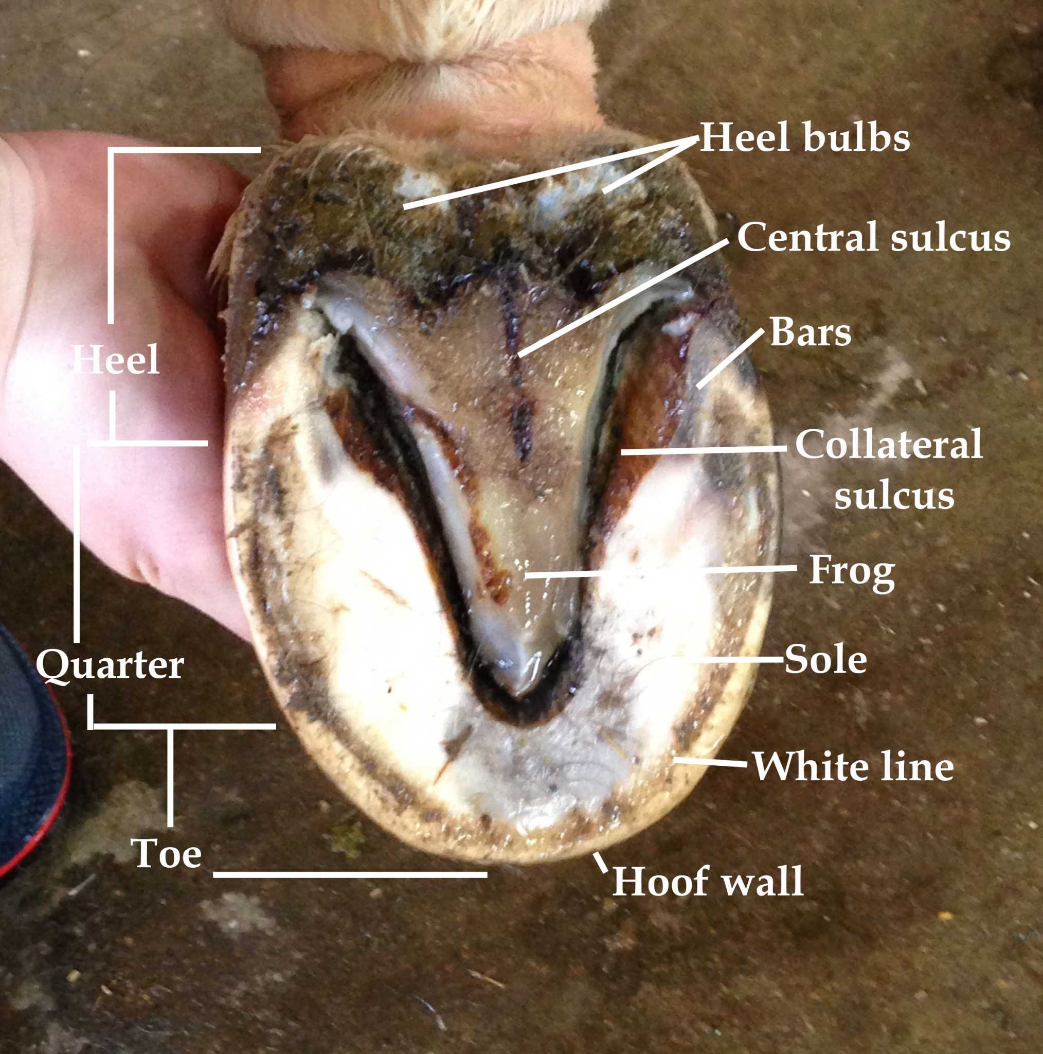
Healthy hoof horn is resilient and alive, expanding at the heels by much as ¼ inch with each step. The heels dampen energy while the frog and sole support the inner hoof tissues, and the toe propels the horse forward. With each foot impact, the hoof pumps blood circulation through the hoof and up the limb. The collateral cartilages are located.

Horn & hoof
The horse's hoof is a miracle of engineering. It contains a whole host of structures which, when healthy, operate in equilibrium with each other to form a hoof capsule which is able to withstand huge forces, utilising energy to assist with forward movement while providing protection to the sensitive structures beneath.

The Anatomy of the Hoof Hoofcount
The hoof is composed of horn, derived from epidermal tissue which has been keratinised to a varying extent . Horn is largely arranged into a series of parallel microscopic tubules, interconnected by intertubular horn [ 9 ].
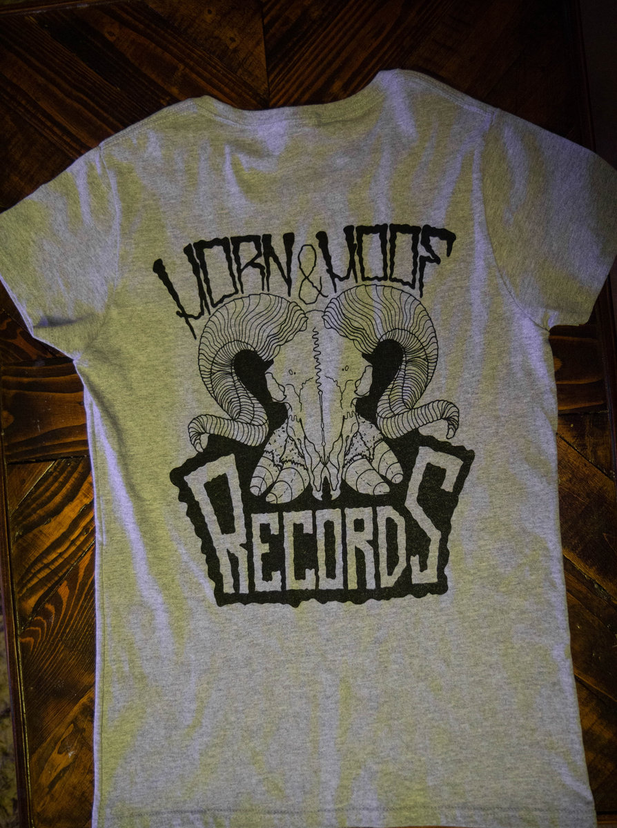
Horn & Hoof shirt (Front and back print) Horn & Hoof Records
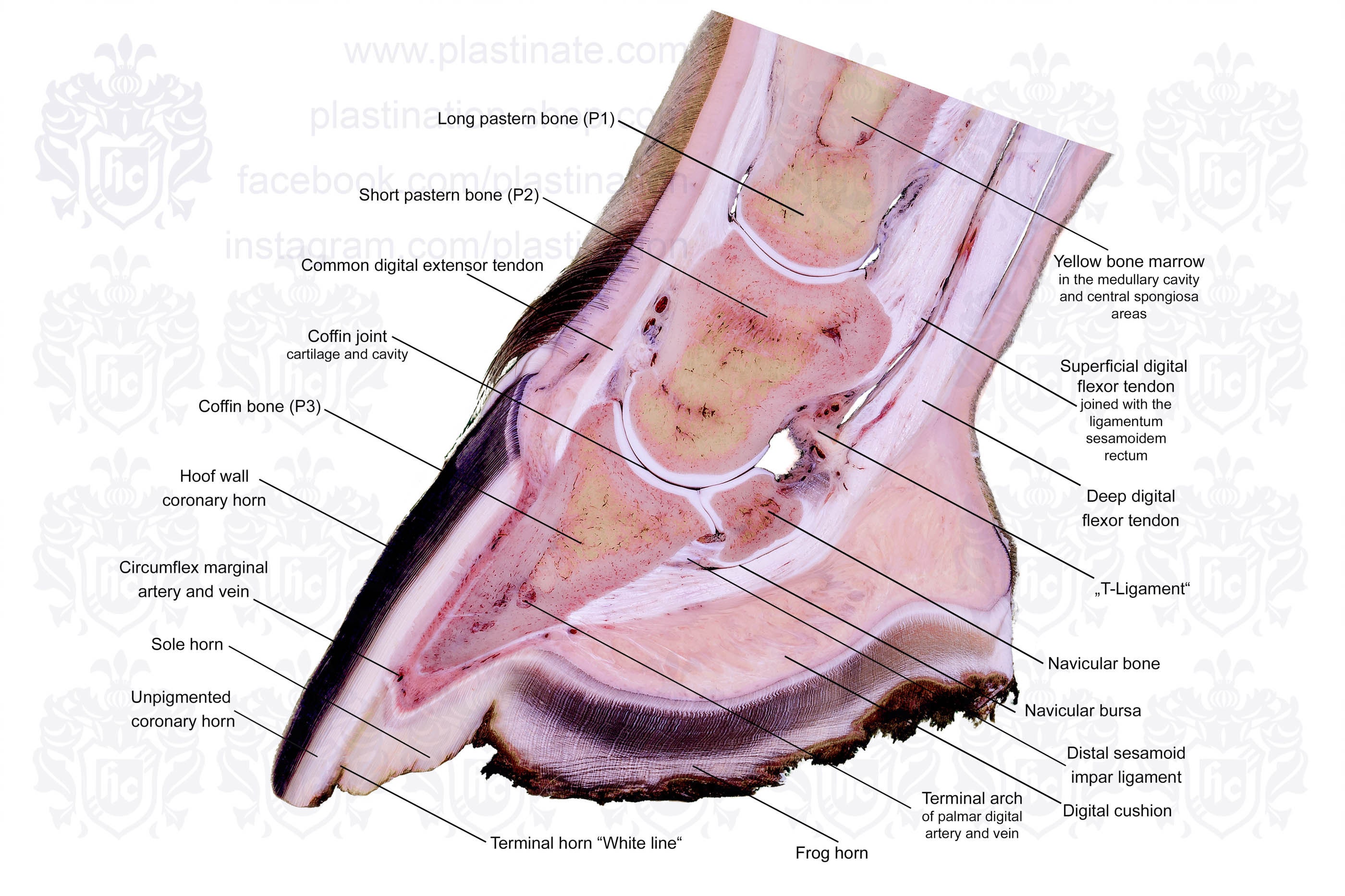
Coffin Bone. The coffin (or "pedal") bone is the bottom bone located near the toe and encapsulated in the hoof. It is the largest bone in the hoof and helps to shape the hoof wall. It's surrounded by special tissues that help make-up the laminae of the hoof wall, as well as, the tissues of the sole.

Hoof Anatomy What Horse Hooves are Made of
Hooves help them walk and run on hard ground. In animals such as the horse and antelope, hooves are an adaptation for fast running and lend the animal both speed and endurance. The sharp hooves of some animals are also used for defense. Hooves are made of keratin, or horn, a fibrous protein that is produced by the outer layer of the skin.

PEELING LUMPS of HOOF HORN from COWS FEET! YouTube
A horn is a permanent pointed projection on the head of various animals that consists of a covering of keratin and other proteins surrounding a core of live bone. Horns are distinct from antlers, which are not permanent.
Outer hoof horn Page 4 Farriers Forum
The hoof is an extremely important structure in an animal's body. Hoof wall is equivalent to our fingernails. This is the strongest horn and is crucial for weight bearing. The Frog is equivalent to the foot pad on a dog or a cat. White line is the junction between the wall horn and sole horn. Its made of weaker horn.
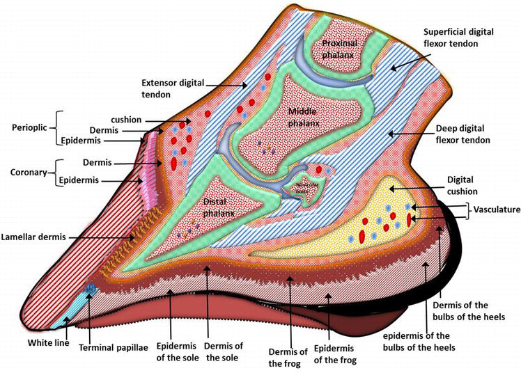
Figure 1.
The horse hoof is the hard covering of the distal end of each digit. If you are a veterinary student, horse physician, or horse owner, you might know the horse hoof anatomy. It is essential for hoof trimming and shoeing purposes. In this short article, I will discuss the horse hoof anatomy with diagrams and natural pictures.

Horn & Hoof Records Resurrect The 'Evil Hoof Picnic'
Hoof horn that is detached from underlying layers of hoof or corium should be removed. Around areas of exposed corium the wall or sole should be thinned to make the existing hoof capsule more flexible along the border of newly developing cornified epithelium. Bandages do not promote healing but may be used to control hemorrhage or to maintain.
Cattle Hoof & Horn
Horn makes up the outer surface if the hoof and is particularly resistant to mechanical and chemical damage. Each epidermal region of the hoof is associated with a dermal region (corium). The corium are connected to the underlying structures by the subcutis.

Horse Anatomy The Hoof The Open Sanctuary Project
A corn is a type of bruise that appears in the sole at the buttress (that is, the angle between the wall and the bar). It is most common in the forefeet on the inner buttress. Corns may arise from pressure applied to the sole by the heel of a shoe improperly placed or left on too long.
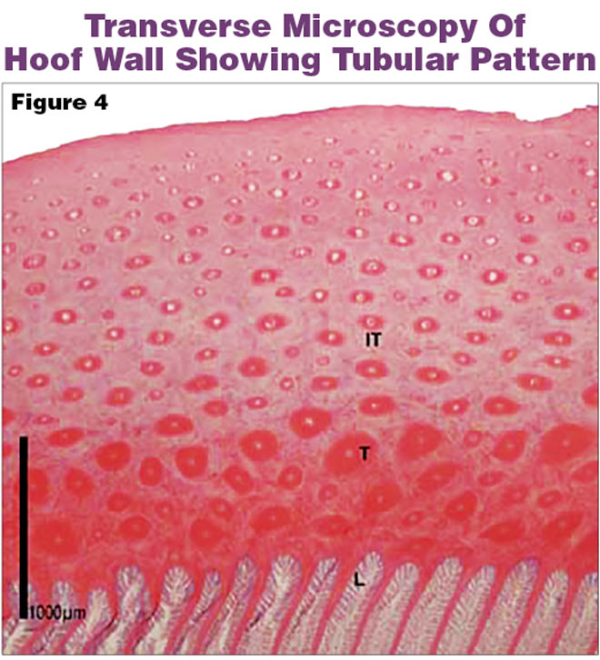
What Do Horn Tubules Do? American Farriers Journal
Although farriers know that horn tubules play an important role in hoof wall structure, this article provides a deeper understanding of it. The equine hoof wall has a complex tubular structure, which extends across the stratum medium. In a healthy hoof, the tubules are straight, parallel to each other and descend at the same angle as the hoof.
Learning about life and death The Rocky Mountain Collegian
The hoof ( pl.: hooves) is the tip of a toe of an ungulate mammal, which is covered and strengthened with a thick and horny keratin covering. [1] Artiodactyls are even-toed ungulates, species whose feet have an even number of digits; the ruminants with two digits are the most numerous, e.g. giraffe, deer, bison, cattle, goat, and sheep. [2]

SheepHoofAnatomy.jpg (800×562) Hooves, Sheep, Goat feet
The distal interphalangeal joint (or DIP joint) is the articulation between the middle and distal phalanges. The ventral aspect of the distal phalanx has the flexor tuberosity, where the deep flexor tendon attaches. Continuous pressure by the flexor tuberosity to the corium may result in hemorrhage and ulceration.

UniqueHorn Hoof Care producten Dierapotheker.be
Founded by Greeks from Syracuse in the 7th century BC, Chiaramonte Gulfi would change both its name and location during the course of its history. It was variously called Akrillai by the Greeks, Acrillae by the Romans, Gulfi ("place of pleasure") by the Arabs, and Chiaramonte for several hundred years. Finally, in 1881 it assumed its present name.
Horse Hoof Anatomy Labelled Teaching Chart Sectional Anatomical Equine Foot Morphology Image
Horn tubules located more peripherally in the hoof wall are smaller (Figure 1) as described by Leach in the mature horse . Table 1 Mean values and (SD) for hoof wall structural variables according to region (dorsal, lateral, medial) of the hooves of 8 foetal Thoroughbreds with ages ranging from 38 days pre-partum to birth.
