Dor sacroilíaca Conheça a sacroileíte Clínica Dr. Hong Jin Pai
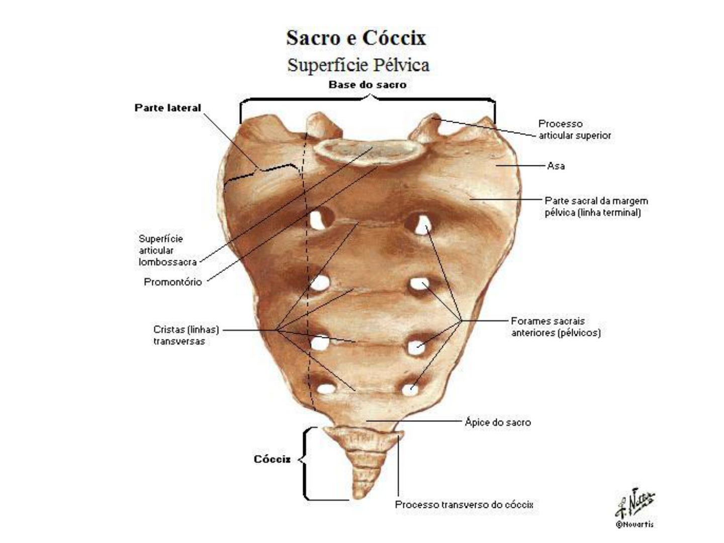
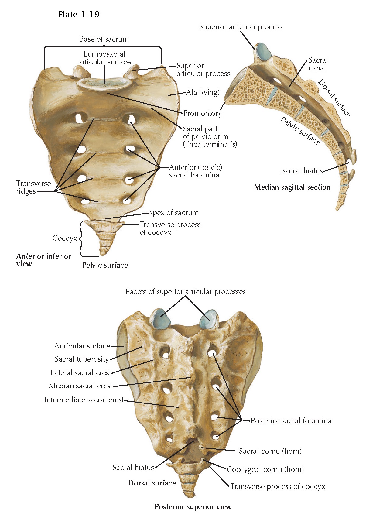
The sacral vertebrae—also called the sacral spine—consists of five sacral vertebrae bones. These bones fuse together to form the sacrum, the shield-shaped bony structure located at the base of the lumbar vertebrae (the five cylindrical bones forming the spine of the lower bank) and connected to the pelvis. The sacral vertebrae are represented by segments S1 through S5 and located between.

Você tem COVINHAS nas COSTAS? Então você está com SORTE!!! Covinhas
"ladder" appearance (15,16). The median sacral vein runs along the midline of the sacrum and is duplicated in up to 80% of patients (12,15,17). In cadaveric studies, the average median sacral vein size has been reported to be 2 mm. The median sacral vein drains into the CIV, almost always on the left, while the LSVs drain into the IIVs.

Sacrum anatomy Sacroiliac joint, Sacroiliac, Lumbar
A covinha ou fosseta sacral é uma pequena depressão na pele logo acima do bumbum do bebê, uma pequena deformidade que ocorre durante o desenvolvimento do bebê no útero. Elas podem ou não indicar uma anomalia na coluna vertebral e acarretar em anormalidades neurológicas e mau funcionamento da bexiga que é controlada pelos nervos dessa região.
Vértebras Sacrais ou Sacro Morfologia
The sacral plexus is a network of nerves emerging from the lower part of the spine. These nerves provide motor control to and receive sensory information from most of the pelvis and leg. A plexus is a web of nerves that share roots, branches, and functions. There are several plexi (plural of plexus) throughout the body, and the sacral plexus.
:max_bytes(150000):strip_icc()/Depositphotos_66748315_original-56a060095f9b58eba4b027de.jpg)
Tilted Pelvis Symptoms, Treatments, Causes and Distinctions
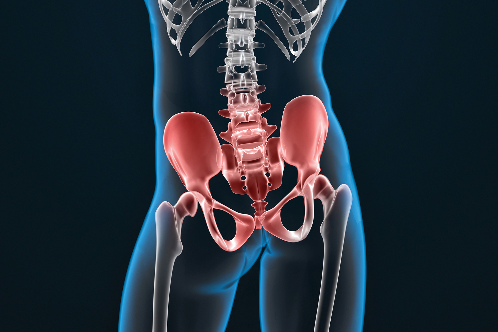
The sacrum is a large trilateral bone located at the base of the vertebral column serving to transfer the body weight from the trunk to the pelvis and lower extremities. Over the years, an abundance of sacral anatomical divergences has been reported, including numerical and/or morphological variations of sacral entities. The majority of these anatomical alternations has been incidentally.
ANATOMY OF THE THORACOLUMBAR AND SACRAL SPINE pediagenosis
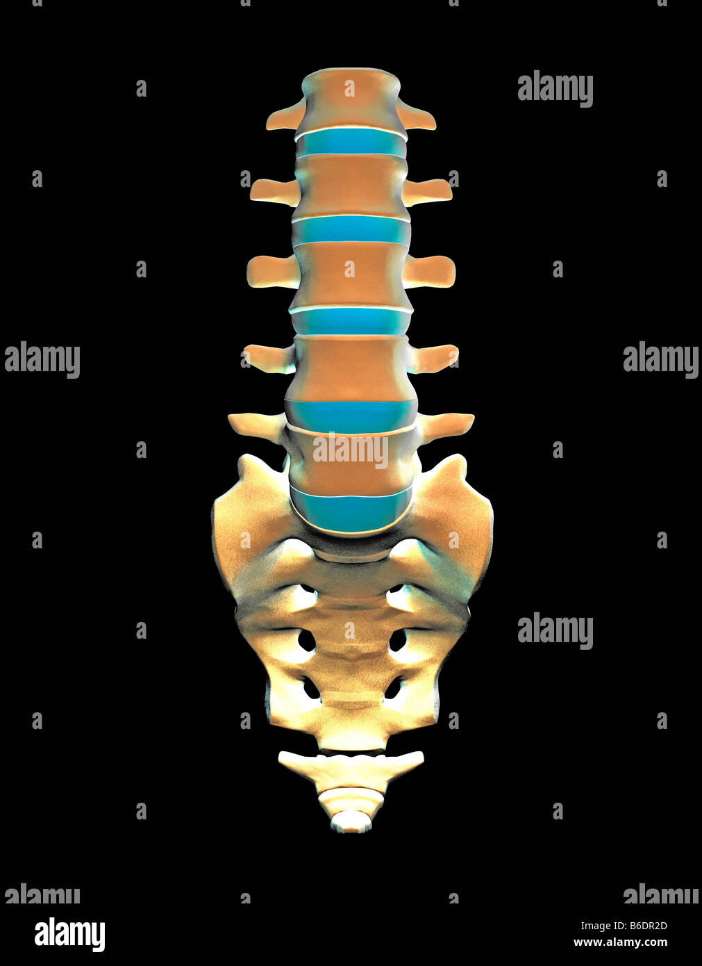
Gross anatomy. The sacrum is an irregularly-shaped bone, shaped roughly like an inverted triangle, with its base superior and apex inferior. It is curved with an anterior concavity and posterior convexity. The sacrum consists of five fused sacral vertebral and costal segments (numbered one-to-five) that form a central sacral body and paired.

Imaging Coccygeal Trauma and Coccydynia RadioGraphics
The cauda equina consists of the spinal nerve roots L2-S5 and the coccygeal nerve.It lies within the distal third of the vertebral canal and extends into the sacral canal. It occupies the lumbar cistern, which is an enlargement of the subarachnoid space containing cerebrospinal fluid (CSF).. Also extending distally from the apex of the conus medullaris is the filum terminale, a vestigial.

Sacrum and Coccyx Anatomy
Purpose To describe the temporal pattern of the appearance of the S1-Co1 centrum ossification centers (COCs) and provide reference data for the S1-S5 COCs and sacral length at various gestational ages (GAs). Methods Postmortem magnetic resonance imaging (MRI) was performed on 71 fetuses (GA, 17-42 weeks) using the 3D dual-echo steady-state with water excitation T2 sequence in the.
articulacion sacro Fisioterapia Granada
7 minutos. A covinha sacral pode ser uma simples depressão na pele ou estar relacionada a patologias do desenvolvimento em alguns casos. Explicamos tudo que você precisa saber e quais são as suas implicações. Alguns bebês nascem com uma pequena depressão na parte inferior das costas. Essa área também é chamada de região lombar ou.

The sacral plexus. Reproduced with permission from Isaacs RE, Fessler
Sacrum. Bony structures and ligaments of the sacrum and coccyx. The sacrum is an irregularly shaped bone, made up of a group of five fused vertebrae in the area of what is commonly known as the base of the spine. Regarded as the keystone of the human body, the sacrum is important because it forms a link between the spine and the iliac bones.
/GettyImages-87394799-56a05fac5f9b58eba4b0275a.jpg)
Sacroiliac Joint Anatomy and Characteristics
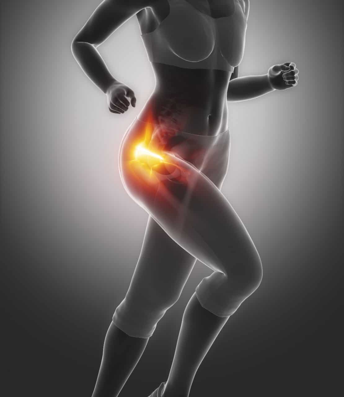
These joints sit where the lower spine and pelvis meet. Sacroiliitis can cause pain and stiffness in the buttocks or lower back, and the pain might go down one or both legs. Standing or sitting for a long time or climbing stairs can make the pain worse. Sacroiliitis can be hard to diagnose. It can be mistaken for other causes of low back pain.

Aula vi.coluna vertebral . part. i
Nos recém nascidos, é importante dar uma atenção especial a qualquer deformidade na região sacral, ou seja, na base da coluna vertebral. Algumas vezes, a criança nasce com anormalidades na pele dessa região. Uma das mais comuns é a presença de um "furinho" ou "covinha". Também há casos em que a criança apresenta manchas.

Sacrum and Coccyx Anatomy
Home treatments for sacroiliitis pain include: Pain relievers you can get without a prescription. Medicines such as ibuprofen (Advil, Motrin IB, others) and acetaminophen (Tylenol, others) may help relieve the pain of sacroiliitis. Some of these medicines can cause stomach upset, or kidney or liver problems. Rest.

Sacrum Bone Anatomy pain — Stock Photo © decade3d 64689121
This is called the sacral canal and the gap inferior to it the sacral hiatus2. The sacral hiatus is normally closed by fibrous tissue that forms the superficial sacrococcygeal ligament. On either side, it has the sacral cornua, which are an extension of inferior processes of the fifth sacral vertebra. These cornua are used to mark the area of.
Lumbar spine and sacrum, The bones of spine (vertebrae) are cream and
Anatomy . The coccyx forms the terminal aspect of the vertebral column. Its name originated from the Greek word for 'cuckoo', as its triangular shape is thought to resemble that of a bird's beak. 2, 3 The dorsal convex aspect of the coccyx has multiple paired coccygeal articular processes, and the most superior of these are termed the coccygeal cornu, which articulate with the sacral cornu.
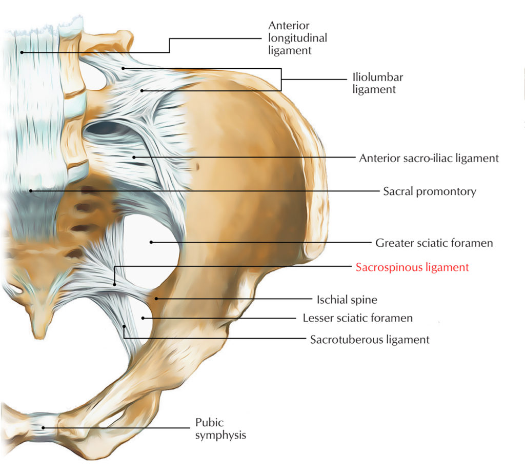
Sacrospinous Ligament Earth's Lab
Uma covinha sacral pode estar localizada no sulco entre as nádegas. No entanto, alguns atributos podem sinalizar mais defeitos, e eles precisarão ser examinados com um ultrassom. Esses incluem: inchaço na área. marcas na pele. uma marca de nascença na área. um pedaço de cabelo pela covinha. um nódulo gordo.
