
Kidney Nephron Model Labeled Medical things Kidney anatomy, Human
Can you Label the Nephron? By LebHead 20m 11 Questions 657 Plays - - Ratings hide this ad Forced Order PLAY QUIZ Score 0/11 Timer 20:00 Quiz Playlist Details Report Classic: Type in answers that appear in a list Forced Order: Answers have to be entered in order Last Updated: Jan 11, 2022 Quiz Scoreboard Sign Up to Join the Scoreboard
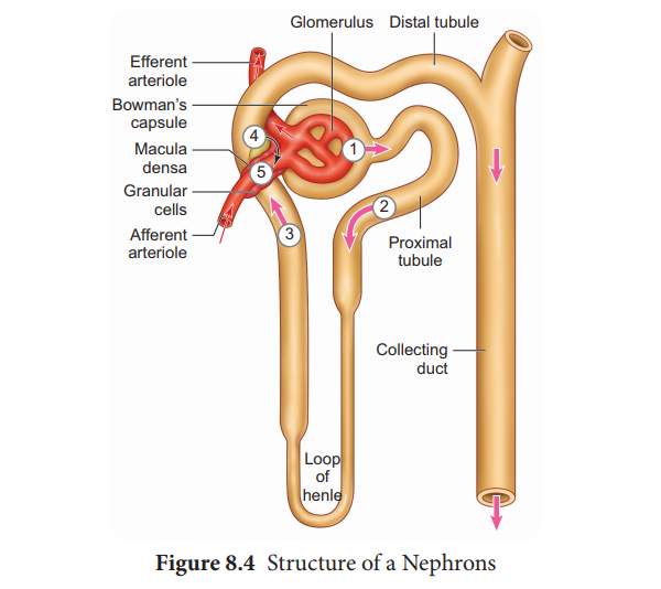
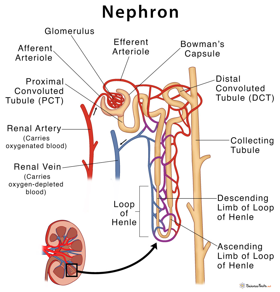
Structure of a nephron
2. The "Bowman's capsule" is the part of a nephron which receives the filtrate. It is a part of a nephron, and only delivers filtrate to a single nephron. The afferent artery and efferent artery are not nephrons, they are arteries outside the nephron that run around the kidney (the red lines that run around the large kidney diagram to the right.
Urinary System Labeling
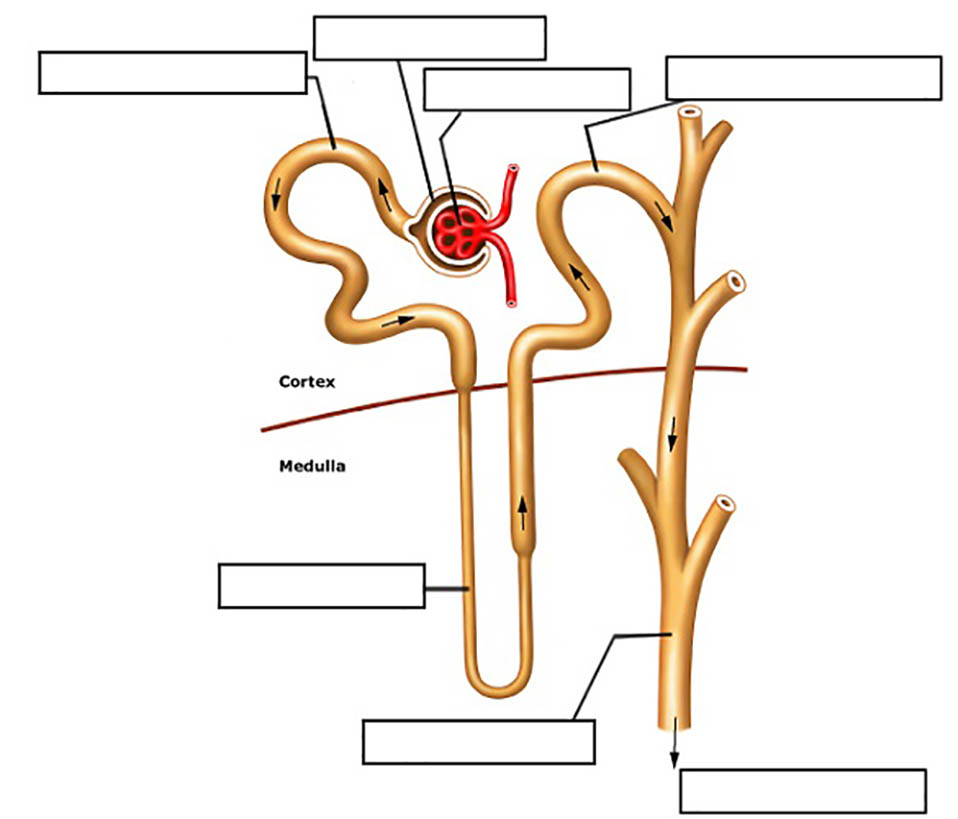
Practice labeling the nephron with this reinforcement activity. Students can also color the image to identify the major structures of the nephron: glomerulus, bowman's capsule, proximal and distal tubules, loop of Henle, collecting duct and capillaries.

Simple Diagram Of Nephron
nephron, functional unit of the kidney, the structure that actually produces urine in the process of removing waste and excess substances from the blood. There are about 1,000,000 nephrons in each human kidney.

The Nephrons Medical Terminology 78 Steps Health
The nephron has three major parts: the glomerulus, the Bowman's Capsule, and the tubules, which consist of the promimal and distal tubule and the Loop of Henle. Blood enters the kidney from the renal artery and moves into the glomerulus, where filtration occurs. Filtration is the process by which water and dissolved particles are pulled out of.
Nephron The Functioning Unit of The Kidney Interactive Biology, with
By correctly labeling the nephron diagram, you will gain a deeper understanding of the complex processes that occur within the kidney and the role that each component plays in maintaining overall health. Structure of a nephron. The nephron is the functional unit of the kidney, responsible for filtering and regulating blood to produce urine.

Diagram of the nephron segments and its juxtaglomerular apparatus
A nephron is the basic unit of structure in the kidney. A nephron is used separate to water, ions and small molecules from the blood, filter out wastes and toxins, and return needed molecules to the blood. The nephron functions through ultrafiltration.
Structure and Functions of Nephron
1/4 Synonyms: Cortex renalis The kidneys are paired retroperitoneal organs of the urinary system. Their function is to filter blood and produce urine. Each kidney consists of a cortex, medulla and calyces. The nephron is the main functional unit of the kidney, in charge of removing metabolic waste and excess water from the blood.
Nephron Definition, Parts, Structure, & Functions, with Diagram
What is Nephron? A nephron is the basic structural and functional unit of the kidney. They are the microscopic structure composed of a renal corpuscle and a renal tubule. The word nephron is derived from the Greek word - nephros, meaning kidney. There are about millions of nephrons in each human kidney. Structure of Nephron
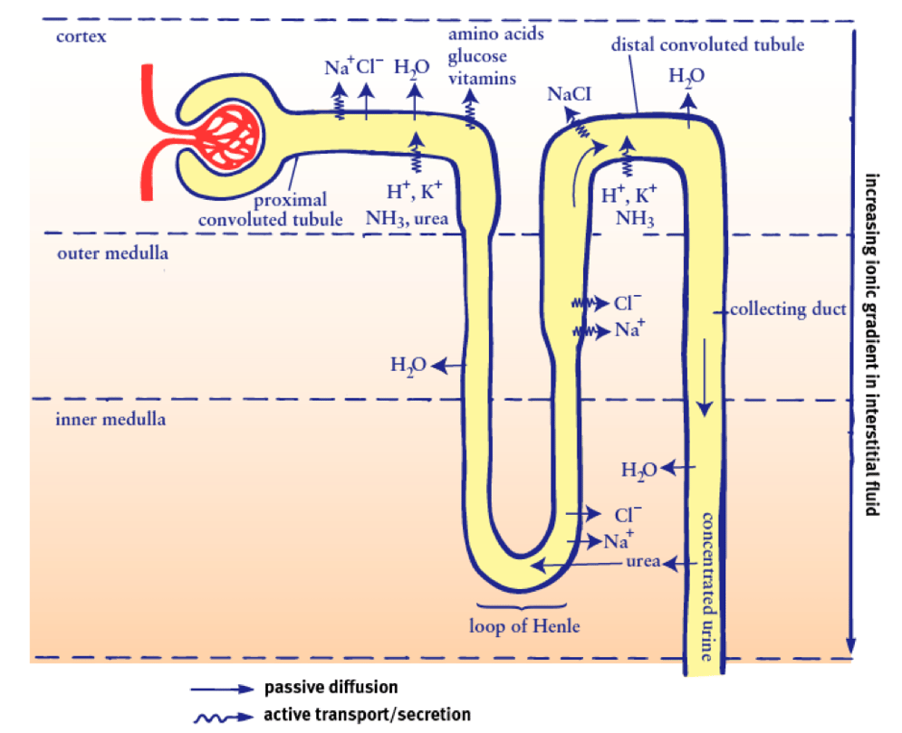
CC Breaking down nephron functioning into six easy steps!
The nephron is the functional unit of the kidney. The glomerulus and convoluted tubules are located in the kidney cortex, while collecting ducts are located in the pyramids of the medulla. (credit: modification of work by NIDDK) Tubular parts of a Nephron - converts the filtrate into urine The Bowman's capsule / Glomerular capsule:

Microscopic Anatomy of the Nephron Medical Pinterest The o'jays
April 5, 2021 in Anatomy, Worksheets by Shannan Muskopf anatomy, kidney, label, learn, nephron, practice, urinary Students practice labeling the urinary system with this drag and drop activity. Three slides have detailed images of the kidneys, ureters, and nephrons.
Nephron of the Kidney Illustration Poster Print by Monica
Describe the composition of urine. Label structures of the urinary system. Characterize the roles of each of the parts of the urinary system. Illustrate the macroscopic and microscopic structures of the kidney. Trace the flow of blood through the kidney. Outline how blood is filtered in the kidney nephron. Provide symptoms of kidney failure.

Kidney Function and Physiology Biology for Majors II
Description. Image of a close up nephron and its place in the kidney. Labels on the kidney cross section show where unfiltered blood enters, filtered blood leaves, and urine exits. On the nephron, the glomerulus, tubule, and collecting duct are labeled along with where unfiltered blood enters, filtered blood exits, and urine exits.

Associate Degree Nursing Physiology Review Human anatomy and
Label a Nephron by ninalahoti 129,291 plays 16 questions ~40 sec English 16p 72 4.36 (you: not rated) Tries Unlimited [?] Last Played December 5, 2023 - 02:42 PM There is a printable worksheet available for download here so you can take the quiz with pen and paper. From the quiz author This is a nephron. Label it. Remaining 0 Correct 0 Wrong 0

Simple Structure Of Nephron Diagram bmpmongoose
A nephron is the unit of structure and function in the kidney. Each nephron is a coiled tube held together by a tough fibrous connective tissue. In humans, a healthy adult has 1 to 1.5 million nephrons in each kidney, functioning together to filter blood from all its impurities.

Kidney Microanatomy (Lesson) Human Bio Media
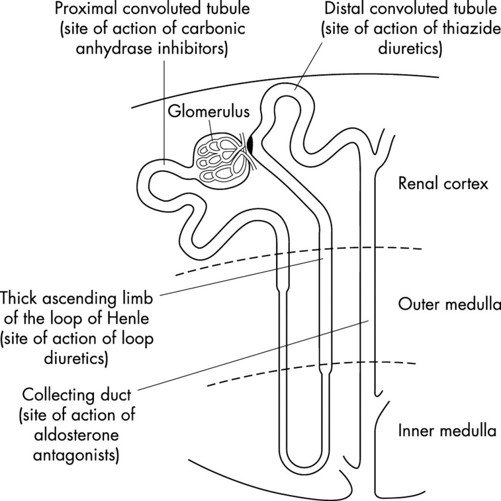
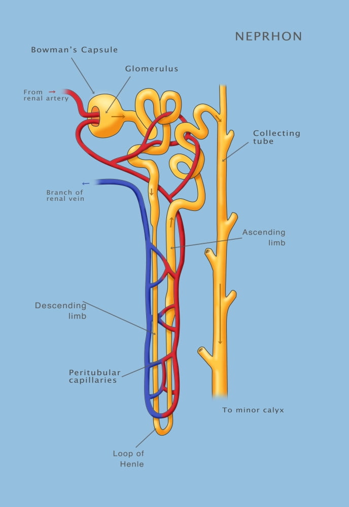
Structure Fig.1) Schematic diagram of the nephron (yellow), relevant circulation (red/blue), and the four methods of altering the filtrate. The nephron is the functional unit of the kidney. [2] This means that each separate nephron is where the main work of the kidney is performed. A nephron is made of two parts:
