Beautiful World Onion cells
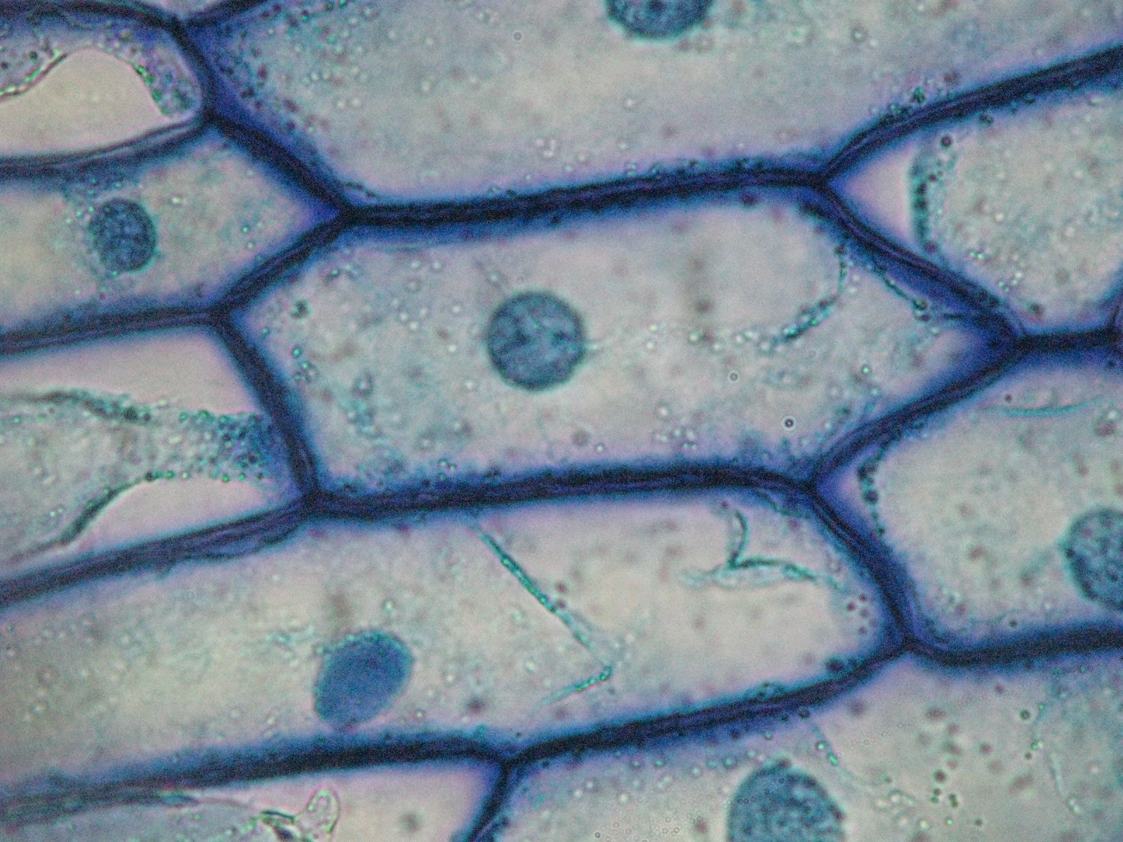
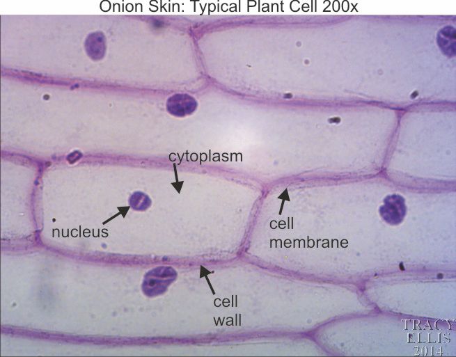
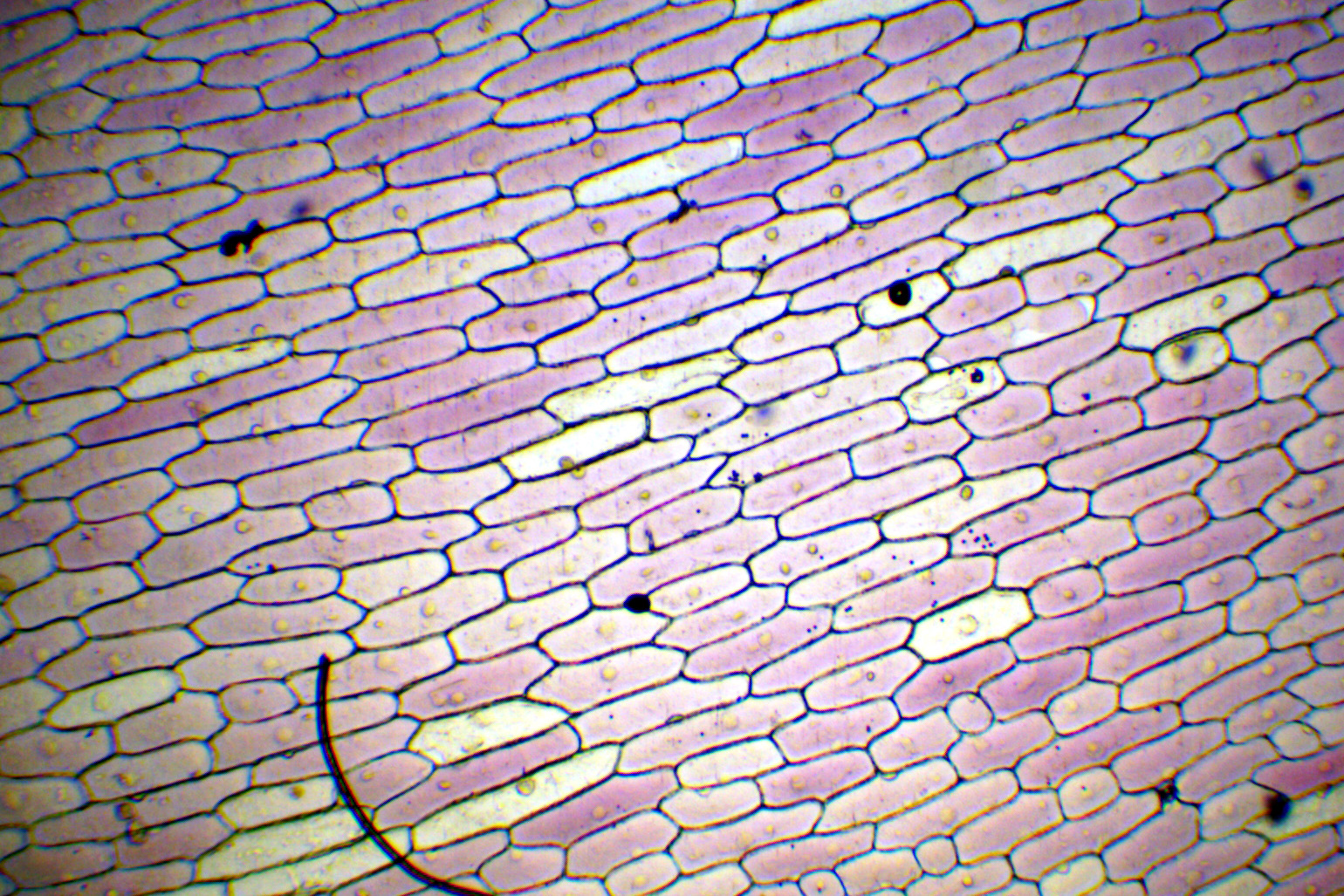
In onion cells the tiles look very similar to rectangular bricks laid in offset runs. The rigid walls combined with water pressure within a cell provide strength and rigidity, giving plants the necessary structure to resist gravity and pressure.
PPT Amoeba PowerPoint Presentation, free download ID6663278
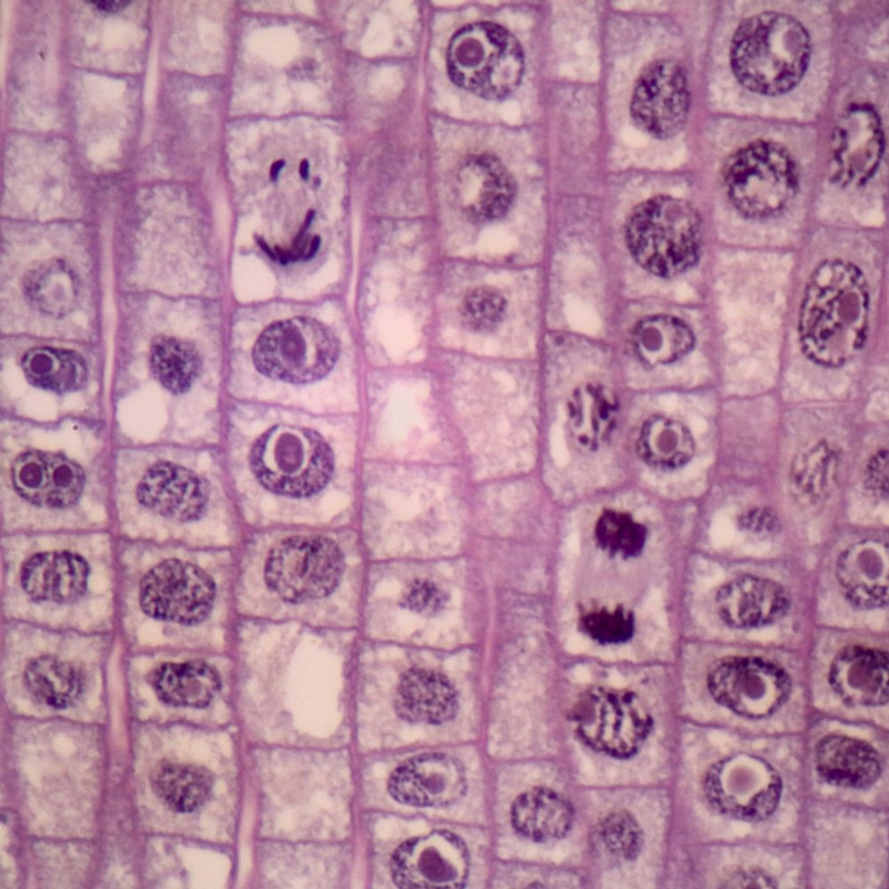
Embed. Introduction to Mitosis in Plant Cells: As a plant cell divides by mitosis, the nucleus, DNA, and mitotic spindle apparatus of a cell follow a specific sequence of events to ensure that a cell's DNA is passed on equally to both daughter cells. Although mitosis is a continual process, scientists have designated several phases (or stages.

draw the figure of an onion peel showing cell Brainly.in
There were three mini-lab procedures carried out during this lab. The first lab exercise was observing animal cells, in this case, my cheek cells. The second lab exercise was observing plant cells, in this case, onion epidermis. The third lab exercise was observing chloroplasts and biological crystals, in this case, a thin section from the.
Onion Mitosis, l.s. Thin Microscope Slide Southern Biological
Onion Wet Mount: Get a clean glass slide and cover slip. Obtain a piece of onion. Use your fingers (nails work well), or forceps, to carefully peel off a small piece of skin from the inner or concave side of the onion chunk. This piece should be thin and translucent, looking much like a piece of scotch tape.
Onion Plant Cell Under Microscope Labeled / Onion Cells Onion epidermis with pigmented large
Procedures: 1. Use the eye dropper to put a drop of water on the slide (will help to flatten out onion tissue). 2. With the tweezers, carefully peel the tissue thin sheet of cells lining the inside of one of the onion layers. 3. Lay the onion tissue gently on the slide (without wrinkling it). 4.
Onion Cell Under Microscope Labeled Drawing apostolicavideo
Figure 10.3.1.1 10.3.1. 1: Cells in an onion root in interphase and prophase. Cell A has a large, dark nucleolus surrounded by greyish material (chromatin) that is enclosed within the nuclear membrane. A cell wall makes a box around each cell and the plasma membrane would be located just inside this box, though we cannot easily see it.

Onion Cell Under Microscope Labeled Drawing apostolicavideo
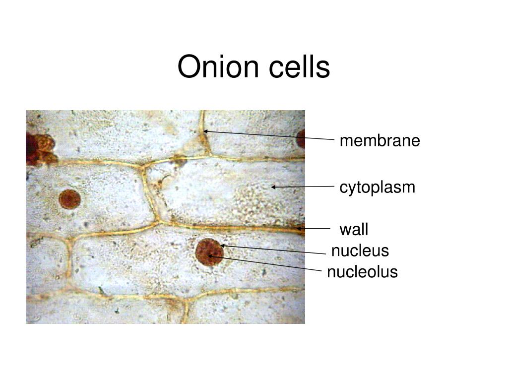
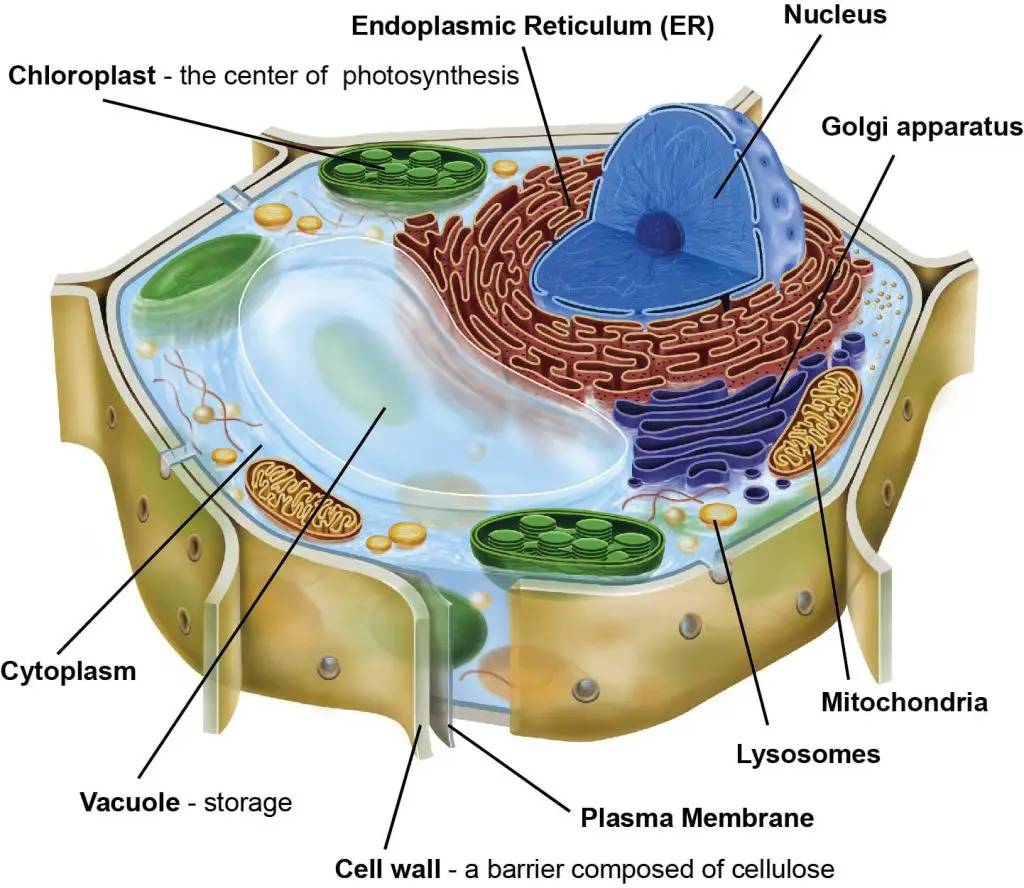
To answer your question, onion cells (you usually use epithelial cells for this experiment) are 'normal' cells with all of the 'normal' organelles: nucleus, cytoplasm, cell wall and membrane, mitochondria, ribosomes, rough and smooth endoplasmic reticulum, centrioles, Golgi body and vacuoles.

Epidermal onion cells under a microscope. Plant cells appear polygonal from the Cell diagram
In general, mitosis occurs through several stages that include: Prophase (divided into prophase and prometaphase) Metaphase. Anaphase. Telophase. Because of the rapid rate at which onion root tips grow as a result of rapid cell division, it's possible to observe and identify the different stages of mitosis.
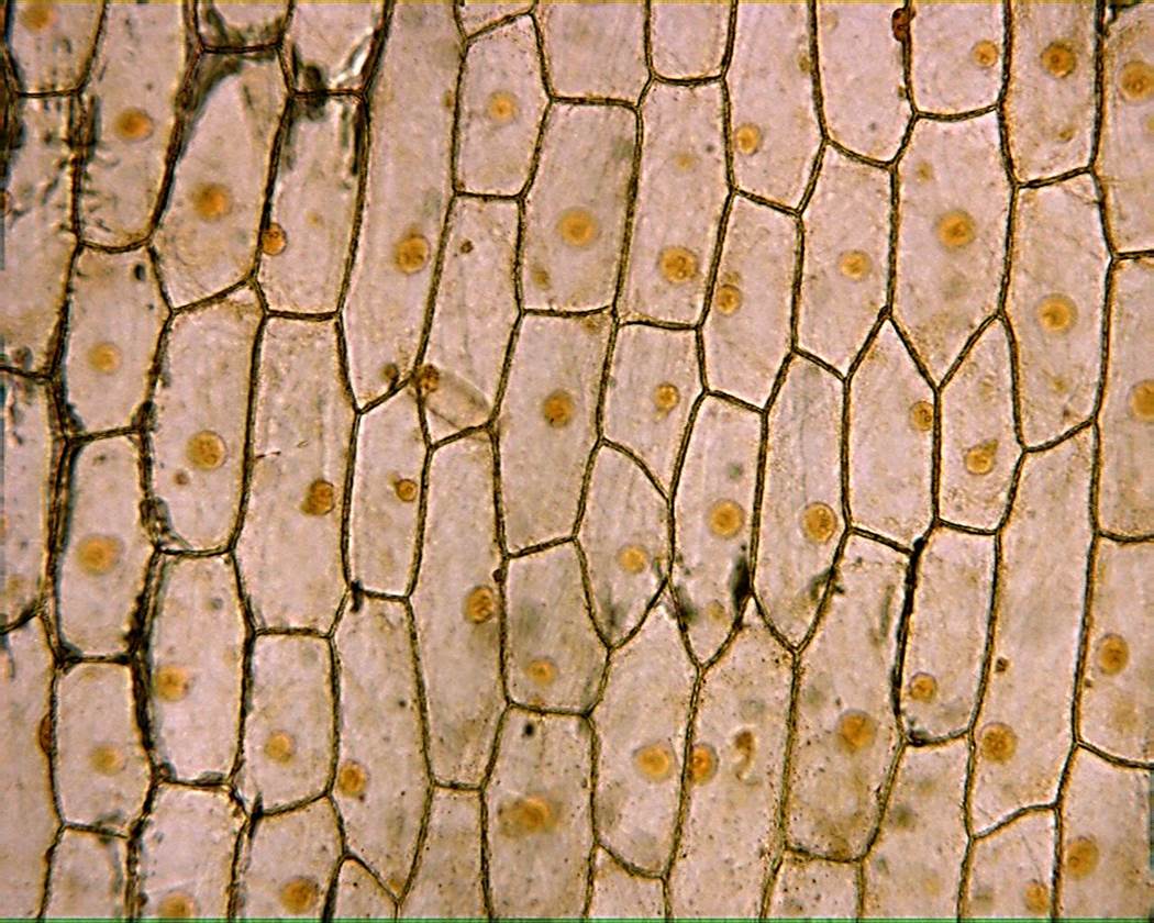
Onion skin 200x Dissection Connection
The Onion Peel Cell Experiment is a popular and educational activity used to observe and understand the structure of plant cells. This experiment focuses on the onion, a eukaryotic plant known for its multicellular composition. As we delve into this experiment, we explore the essential components that make up a cell, the building blocks of life.

The layer present over the cell membrane in an onion cell is called
Mitosis in an Onion Cell This graphic shows an image of what cells in an onion root tip would look like as they are in various stages of mitosis. This worksheet compliments a laboratory activity where students look at onion root tip slides to identify phases of mitosis.

onion cell diagram Laceness
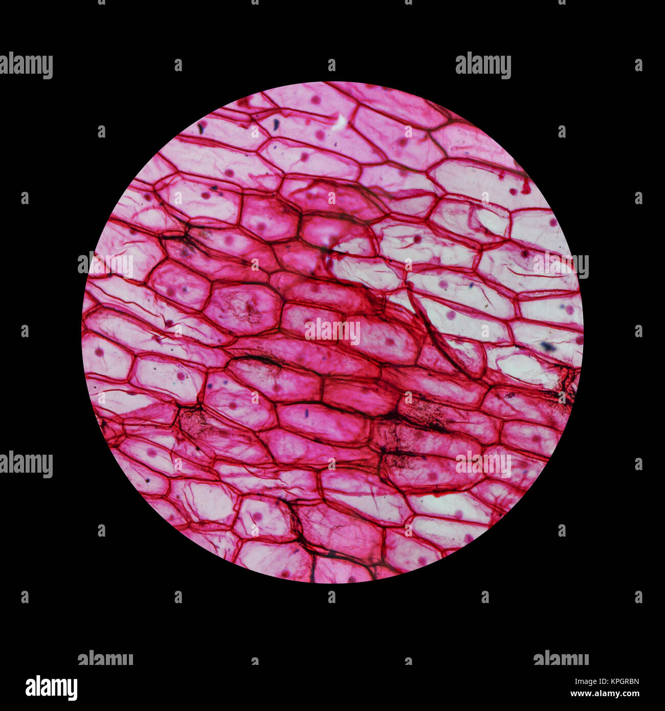
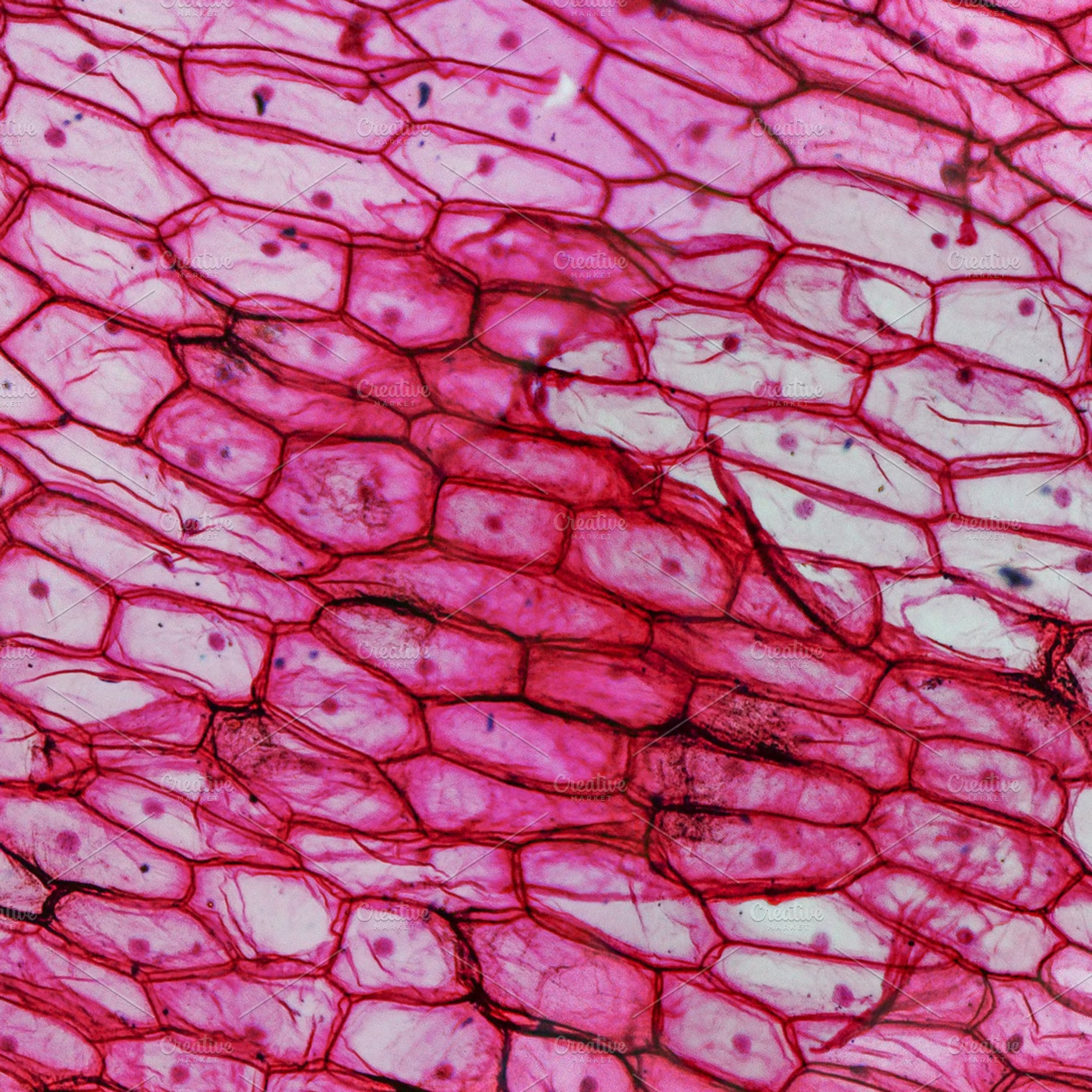
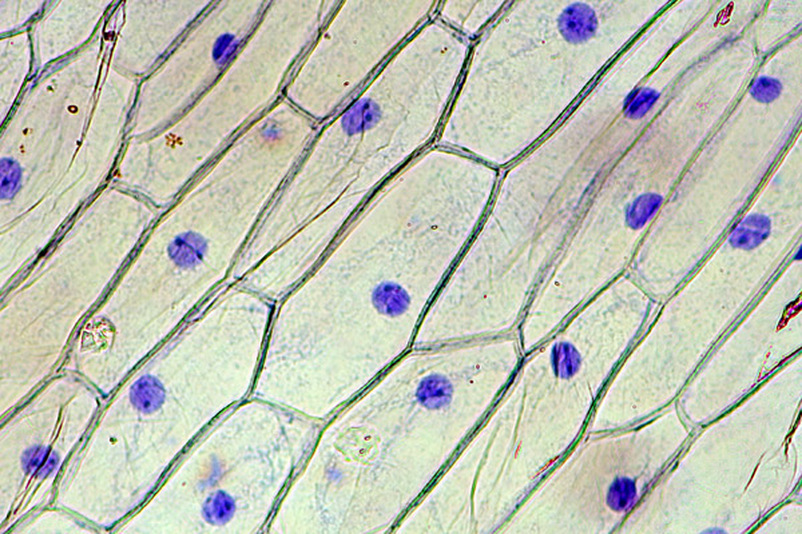
Onion epidermal cell These large cells from the epidermis of a red onion are naturally pigmented. The epidermal cells of onions provide a protective layer against viruses and fungi that may harm the sensitive tissues.
Onion cells HighQuality Nature Stock Photos Creative Market
An onion is a multicellular (consisting of many cells) plant organism. As in all plant cells, the cell of an onion peel consists of a cell wall, cell membrane, cytoplasm, nucleus and a large vacuole. The nucleus is present at the periphery of the cytoplasm. The vacuole is prominent and present at the centre of the cell.
Onion Epidermis 100X General Biology Lab Loyola University Chicago
Onion Cell Lab Power __________ Total Magnification __________ After you have completed the rest of this lab come back to this cover page DRAW & LABEL AN ONION CELL WITH ALL THE PARTS / ORGANELLES YOU OBSERVE UNDER 40X. Purpose: To observe and identify major plant cell structures and to relate the structure of the cell to its function.
Microscopy
Label the cell wall, middle lamella, plasmodesmata, and chromoplasts. You are encouraged to identify and label other cell components, such as the nucleus and nucleolus, if they are visible. A potato is a modified part of the plant called a tuber. Much like an onion, a tuber is a part of the plant--this time the stem--adapted for storing starch.
Onion Cell
These regions of growth are good for studying the cell cycle because at any given time, you can find cells that are undergoing mitosis. In order to examine cells in the tip of an onion root, a thin slice of the root is placed onto a microscope slide and stained so the chromosomes will be visible. The cells you'll be looking at in this activity.
Onion Plant Cell Under Microscope Labeled / Onion Cells Onion epidermis with pigmented large
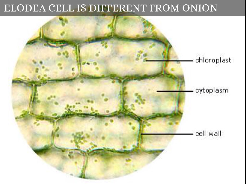
Onion Cells Under a Microscope ** Requirements, Preparation and Observation The bulb of an onion is formed from modified leaves. While photosynthesis takes place in the leaves of an onion containing chloroplast, the little glucose that is produced from this process is converted in to starch (starch granules) and stored in the bulb.
