
Snake X Ray Royalty Free Stock Photos Image 21038838
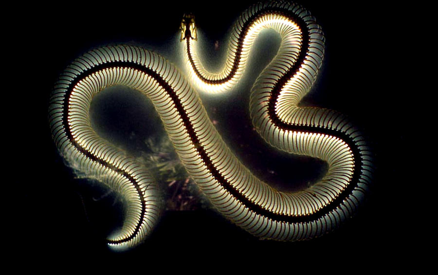
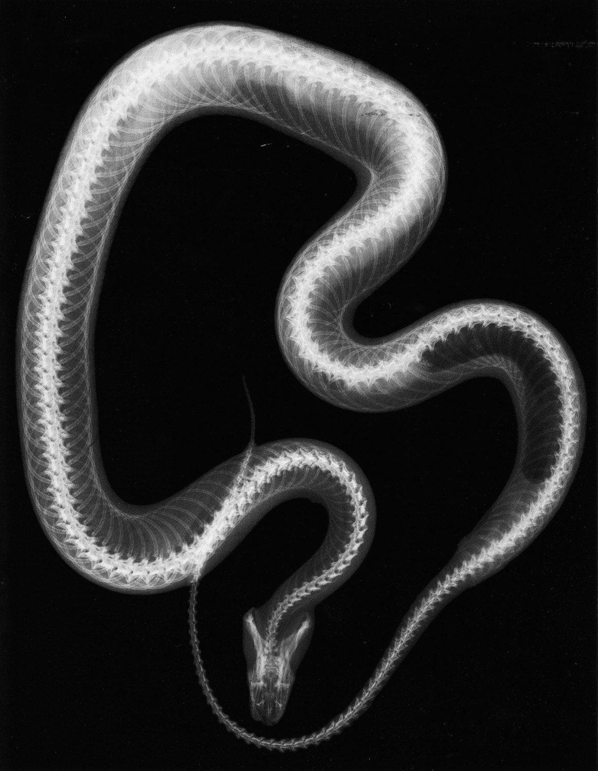
Caption. Snake, coloured X-ray. The snake's skull is at bottom right, attached to the top of a long spine. The spine can have between 130 and 500 vertebrae, depending on the species.

Striking Xray pictures show inner workings of London Zoo animals Express & Star
Synchrotron X-ray investigation of a fossilised snake with legs is helping scientists better understand how in the course of evolution snakes have lost their.
Snake XRay by Katzilla13 on DeviantArt
An x-ray image shows a snake that entered a woman's stomach by crawling through her vagina. Rating: False About this rating Origin A purported x-ray image supposedly showing a long.

Snake Xray. This only kind of cool, and really creepy. Plus a little gross... Xray Art, Law Of
Snakes may be constrained manually or encouraged to crawl into a perspex or plastic tube, such as can be made from syringe cases taped together. This has two advantages. One is that the snake is adequately restrained enough to allow the handler to leave the snake whilst the radiograph is being taken.

images of interesting x rays Snake XRays.... Snake, X ray, Ultra violet
Micro-CT imaging is highly effective for the comprehensive study of internal structures and skeletal elements of snakes, and can facilitate morphometric studies, phylogenetics analysis, and biomechanics modeling. In addition to skeletal studies, including research on the head, jaw, and tail, current research also focuses on studying venomous snake fangs for inspiration in developing new.

This xray by Two Vets Talk Pets Podcast revealed what happened to that second snake. Pet
X-Ray Microscopic Imaging of a Rattlesnake This month we imaged a rattlesnake at three scales using the SkyScan 1273 micro-CT. The large sample volume of the SkyScan 1273 allowed us to examine the snake at a full body overview scale and two higher resolution captures of regions of high interest - the head and tail.

Snake Xray Xray art, X ray, Snake
Reptile Radiography Issue: November/December 2014 Danielle Mauragis CVT Clifford R. Berry DVM, DACVR Radiography of reptile patients is routinely used for evaluation of traumatic injuries and the gastrointestinal and reproductive tracts. A reptile radiography study typically includes lateral and dorsoventral views.

Snake, Xray Stock Image C011/6061 Science Photo Library
A new look at a 95-million-year-old fossilized snake reveals two tiny leg bones attached to the slithery creature's pelvis.. (CT) scan, SRCL uses X-rays to image the internal structure of an.
Gravid Ball Python XRay r/snakes
Aug 22, 2022 at 11:09 AM EDT By Kate Fowler Internet Culture & Trends Reporter Zoo Miami made a surprising discovery when it couldn't locate a Burmese python it had been tracking in Florida..
Xray of our corn snake r/snakes
A dorsoventral view can be helpful to identify foreign bodies, intestinal impaction, or coelomic masses. A horizontal x-ray beam provides the best lateral imaging in lizards, especially when evaluating the respiratory system. The positioning for this view involves rotating the x-ray tube 90 degrees and placing the film vertically behind the lizard.

Incredible Xrays show how snake swallowed a doorknob it thought was an egg Daily Mail Online
Snake x ray Stock Photos and Images (143) See snake x ray stock video clips Quick filters: Cut Outs | Vectors | Black & white Sort by Relevant RF 2CCEPKA - Snake, X-ray RM 2BE0J79 - Historical X-ray of an Aesculapian snake, 1896.
Had to get our ball python an xray. Thought this would be interesting to share. r/snakes
A novel X-ray imaging technology is helping scientists better understand how in the course of evolution snakes have lost their legs. The researchers hope the new data will help resolve a heated.

1000+ images about XRay on Pinterest Natural history, Paintball guns and Python snake
The 19-inch-long (50 centimeter) snake (called Eupodophis descouensi) is one of only three snake fossils with its hind limbs preserved, so breaking it open to look for the other leg was out of.
Snake Xray Real xray
X-rays reveal hidden leg of an ancient snake. This is a 3-D reconstruction from synchrotron X-ray images of the previously hidden second leg of Eupodophis. The bones are artificially colored to.

Snake, Xray Stock Image C021/2795 Science Photo Library
This is an X-ray of the two headed snake. You will notice the skulls have a dragon-like look. Our snake was only 56 cm or 22 inches long, but prehistoric snakes could have been much bigger allowing people to imagine large demons. Feeding time required some clever thinking by our animal caretaker since the two heads merge at the neck. To keep the snake from choking one head

Snake, Xray Stock Image C025/8519 Science Photo Library
Often, necessary positioning can be achieved using simple positional aids, such as Perspex tubes for snakes, beakers, Perspex boxes and masking tape. 1 A nifty trick for imaging the limbs of chelonians is to suspend the patient on an upturned bucket or similar structure.
