Physical Therapy in Buffalo for Knee Anatomy
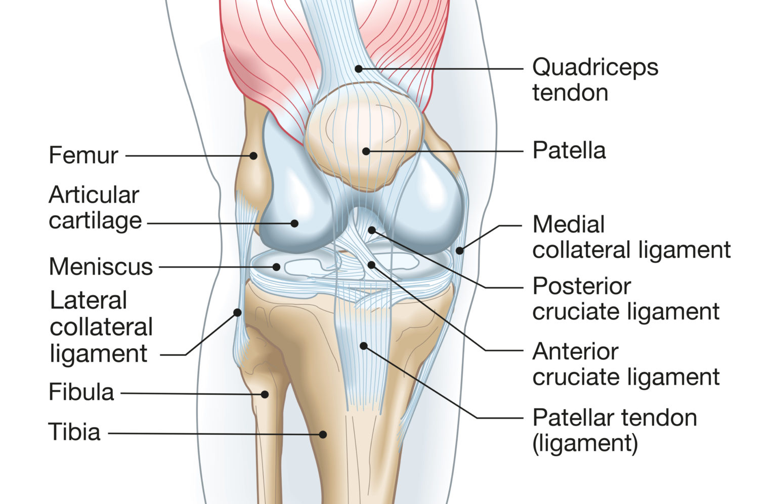
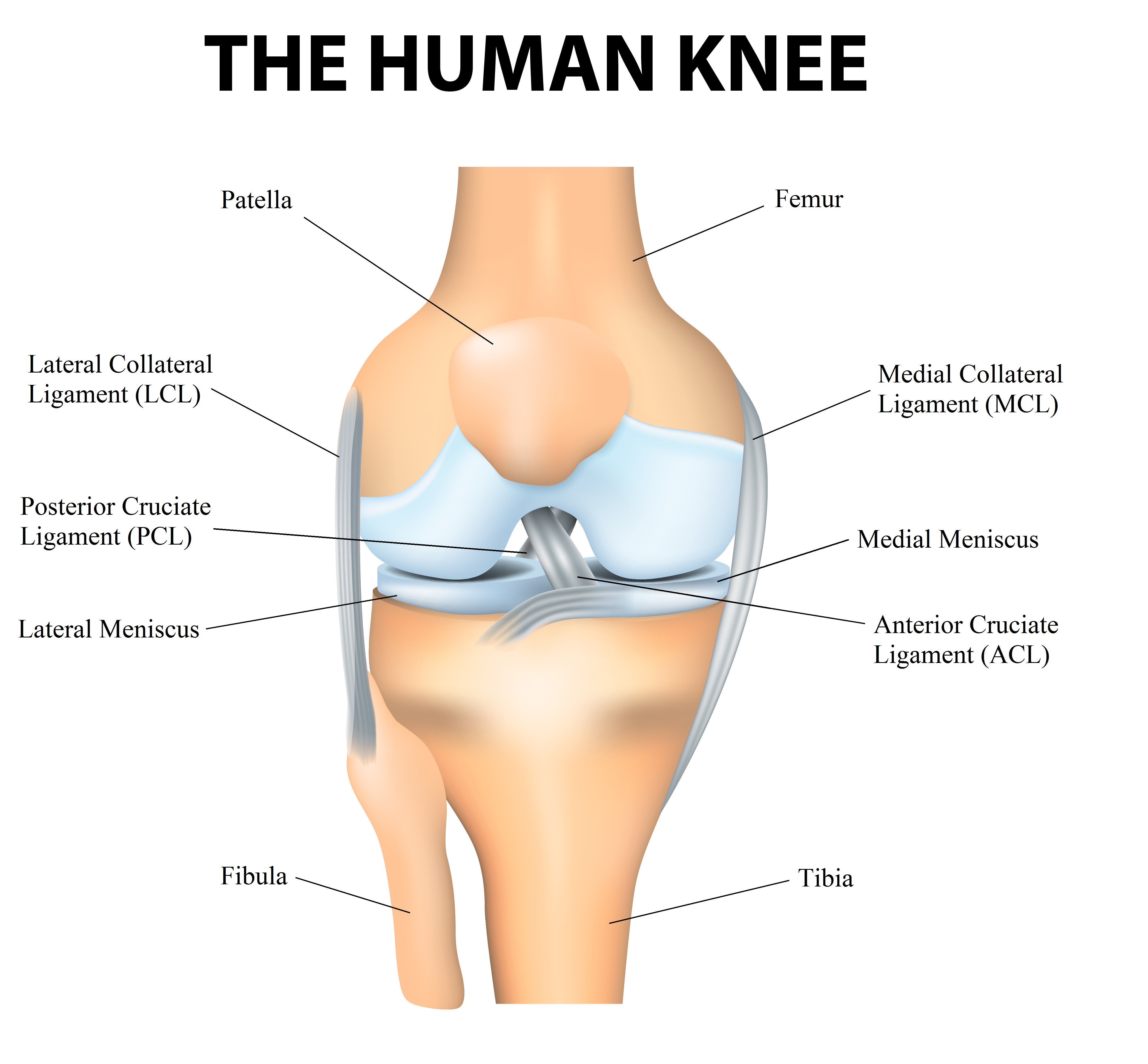
Knee ligaments are bands of tissue that connect the thigh bone in the upper leg to the lower leg bones. There are four major ligaments in the knee: ACL, PCL, MCL and LCL. Injuries to the knee ligaments are common, especially in athletes. A sprained knee can range from mild to severe.

Knee Anatomy
Browse 2,712 knee joint anatomy photos and images available, or search for knee pain to find more great photos and pictures. Browse Getty Images' premium collection of high-quality, authentic Knee Joint Anatomy stock photos, royalty-free images, and pictures.

Sydney Knee Specialists Kogarah, Miranda & Sydney
Tibialis anterior. Tibialis posterior. Tibiofibular joint; Superior tibiofibular joint. Transverse ligament of knee. Vastus intermedius. Vastus lateralis. Vastus medialis. Atlas of the anatomy of the joint of the knee on a CT arthrogram in axial, coronal, and sagittal sections, on a 3D images and on conventional arthrogram.
Knee Pain Causes, Exercises, Remedies, Medication & Treatment
49,887 knee anatomy stock photos, 3D objects, vectors, and illustrations are available royalty-free. See knee anatomy stock video clips Filters All images Photos Vectors Illustrations 3D Objects Sort by Popular Accurate medically illustration showing knee joint with ligaments, meniscus, articular cartilage, femur and tibia. Knee anatomy.
:max_bytes(150000):strip_icc()/knee-anatomy--artwork-452427829-599d8b9b22fa3a0011f2030d.jpg)
What Is Causing Your Knee Pain?
PeopleImages / Getty Images Anatomy . The medial compartment of the knee includes everything within the inner half of the joint and is located where the tibia (shinbone) and femur (thigh bone) meet. The rounded end of the femur bone (medial femoral condyle) sits on a flattened area of the tibia bone called the medial tibial plateau.
Knee Boundless Physical Therapy & Sports Performance
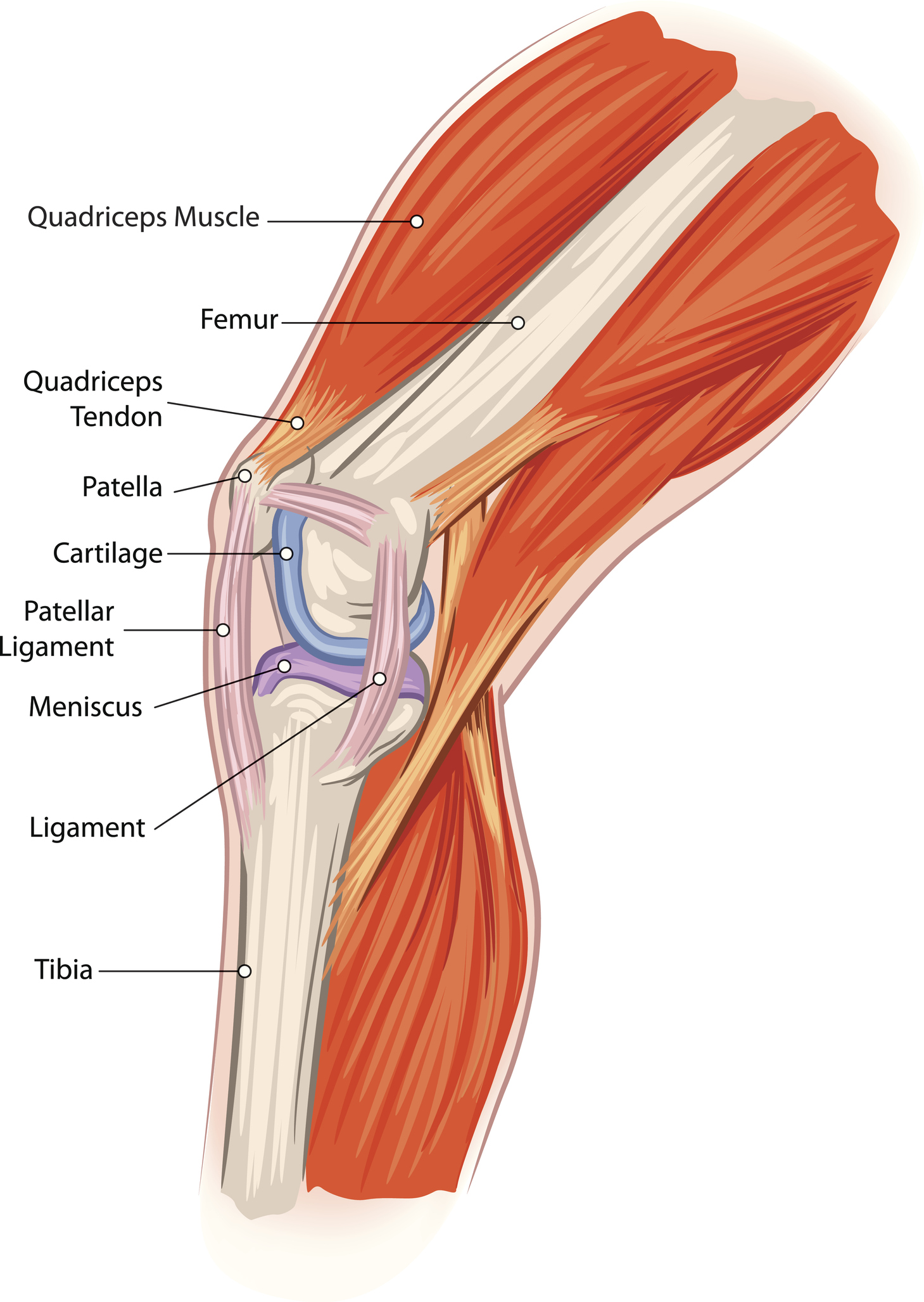
It consists of bones, meniscus, ligaments, and tendons. The knee is designed to fulfill a number of functions: support the body in an upright position without the need for muscles to work. helps.
Knee Pain Causes, Exercises, Remedies, Medication & Treatment
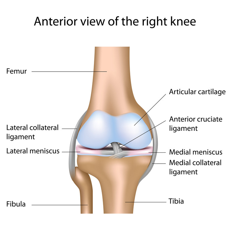
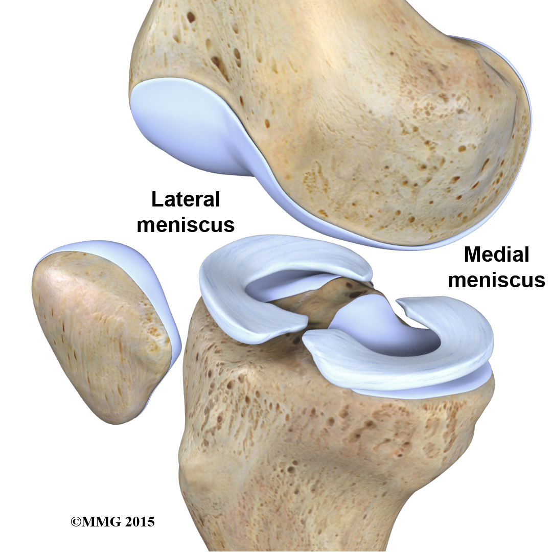
SPRINGER MEDIZIN / Getty Images The knee bones are lined with a smooth cover known as articular cartilage. This slippery substance that help the bones glide as the joint moves. Two wedge-shaped piece of meniscus cartilage sit between the articular cartilage of the femur and tibia.
Common Knee Injuries OrthoInfo AAOS
Knowing about knee anatomy can help people understand how knee arthritis develops and sometimes causes pain. See Possible Causes of Severe Knee Pain. Show Transcript The knee joint is a hinge joint, meaning it allows the leg to extend and bend back and forth with minimal side-to-side motion. It is comprised of bones, cartilage, ligaments.
Anatomy, Pathology & Treatment of the Knee Joint Articles & Advice
This atlas of cross-sectional anatomy of the knee is based on magnetic resonance imaging (MRI). Each anatomical structure was labeled interactively.
Anatomy of the Knee Knee Specialist Fairfield Shelton Stratford
4K HD. of 100 pages. Try also: knee anatomy in images knee anatomy in videos knee anatomy in 3D knee anatomy in Premium. Search from thousands of royalty-free Knee Anatomy stock images and video for your next project. Download royalty-free stock photos, vectors, HD footage and more on Adobe Stock.
FAQs on Knee Injuries Answered by a Los Angeles Knee Surgeon
Browse 1,136 knee anatomy picture photos and images available, or start a new search to explore more photos and images. Browse Getty Images' premium collection of high-quality, authentic Knee Anatomy Picture stock photos, royalty-free images, and pictures.
The Knee UT Health San Antonio
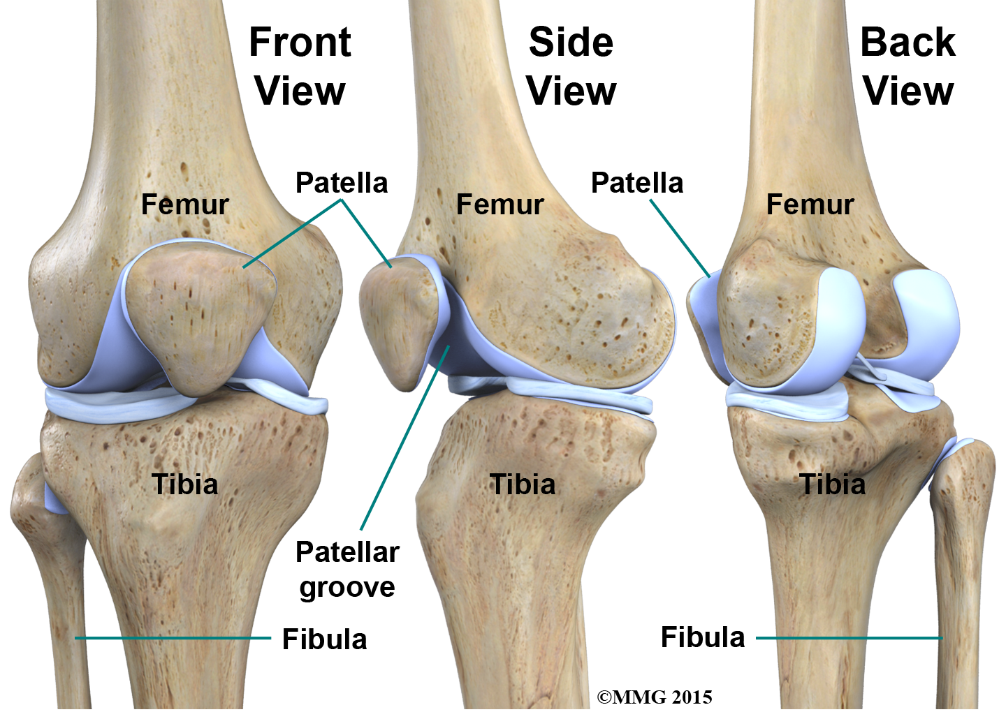
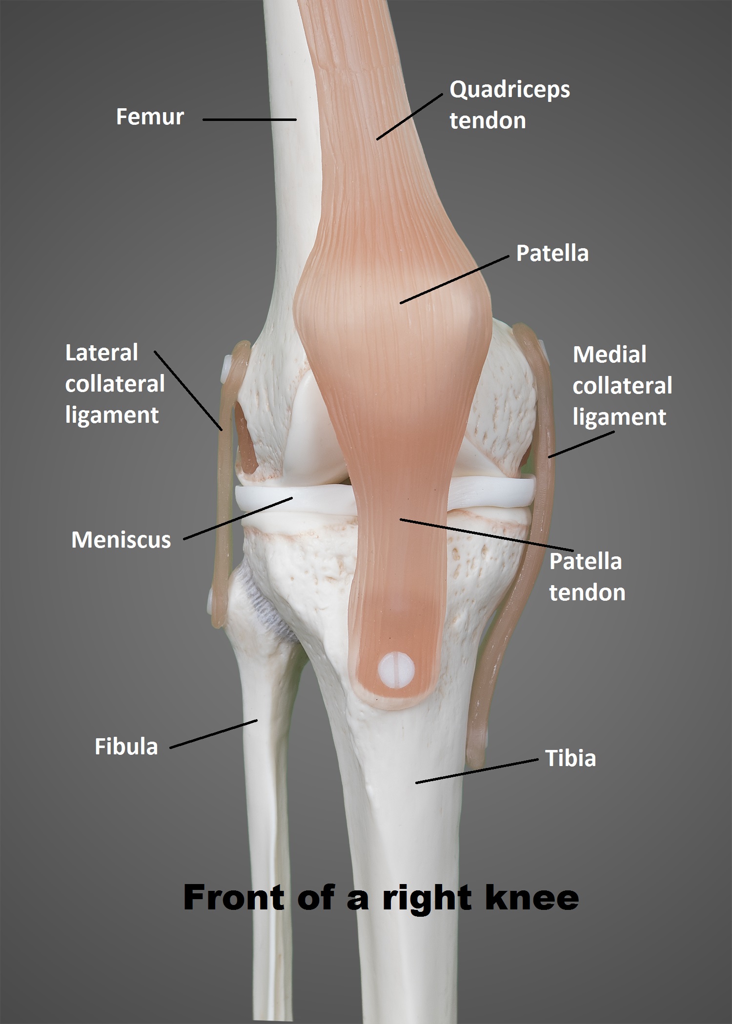
The knee joint is a synovial joint that connects three bones; the femur, tibia and patella. It is a complex hinge joint composed of two articulations; the tibiofemoral joint and patellofemoral joint.
Physical Therapy in Jackson for Knee Anatomy
Function What does the knee joint do? Your knees have several important jobs, including: Moving your legs. Supporting your body when you stand and move. Stabilizing you and helping keep your balance. Anatomy Where is the knee joint located? The knee is the joint in the middle of your leg.

Muscles of the Knee Anatomy Pictures and Information
Browse 18,800+ knee anatomy stock photos and images available, or search for knee anatomy illustration or human knee anatomy to find more great stock photos and pictures. knee anatomy illustration human knee anatomy Sort by: Most popular Knee anatomy Knee anatomy. Structure of leg joint. Major parts.

Knee Anatomy Posterior View Human Anatomy
Knee joint anatomy involves looking at each of the different structures in and around the knee. The knee joint is the largest and one of the most complex joints in the human body.

FileKnee diagram.svg Wikipedia
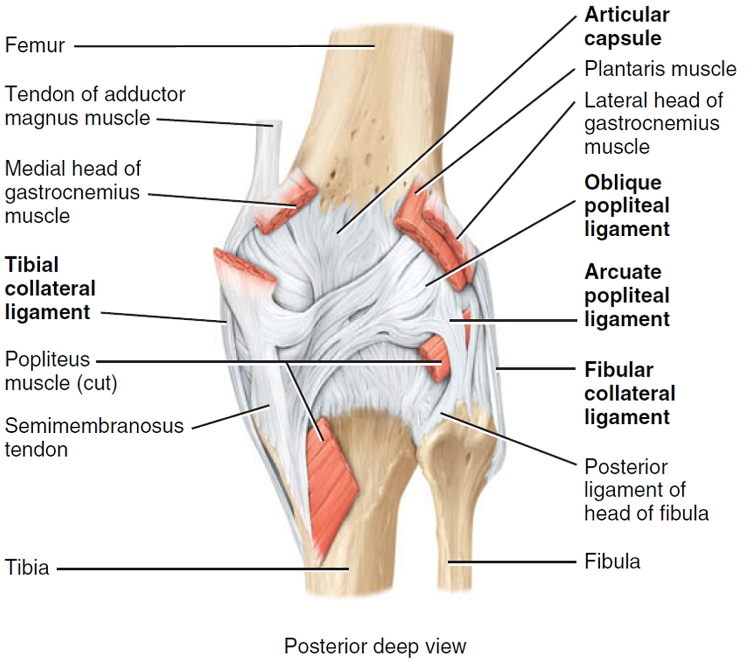
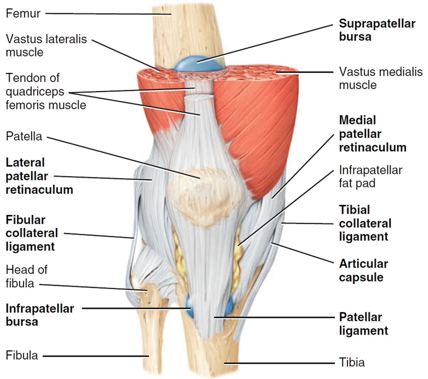
Description. The knee joint is one of the largest and most complex joints in the body. It is constructed by 4 bones and an extensive network of ligaments and muscles. [1] It is a bi-condylar type of synovial joint, which mainly allows for flexion and extension (and a small degree of medial and lateral rotation). [2]
